page 3 >
5e) Explaining ME/CFS? Prusty / Naviaux Study Ties Infections
Explaining ME/CFS? Prusty / Naviaux Study Ties Infections to Energy Breakdownsby Cort Johnson | Apr 26, 2020 | Autoimmune, Energy Production, Homepage, Human Herpes Virus Six (HHV-6), Immune 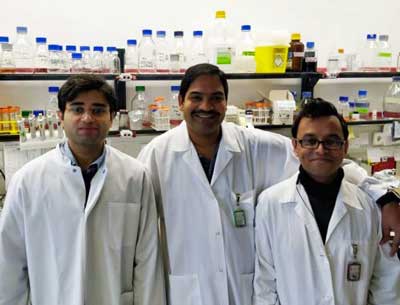 Prusty’s novel work on HHV-6 has been supported by the Solve ME/CFS Initiative and the HHV-6 Foundation. Unexpected synchronies are always a good sign. Many, of course, are familiar with Bob Naviaux, MD, PhD from the University of California, San Diego (UCSD). Naviaux’s metabolomic work and his Cell Danger Response (CDR) hypothesis have opened up new possible ways of understanding ME/CFS, autism and other diseases. Naviaux is best known for his metabolomic work, but what most people don’t know is that Naviaux is also a Salk-trained virologist, who invented some early retroviral gene transfer vectors, and was trained, to boot, in natural killer cell biology – a key topic in ME/CFS. Bhupesh Prusty, PhD of the University of Würzburg in Germany, is newer on the scene but has been raising eyebrows with his proposal that herpesviruses like HHV-6 (and other viruses as well) may be knocking the mitochondria in ME/CFS patients for a loop. The two authors – Prusty and Naviaux were the co-senior authors who conceived the project – teamed together to attempt to answer a question that’s been plaguing patients, doctors and researchers for years: how to tie together the energy problems in ME/CFS with the infectious onset that so many patients experience. Coming from two separate fields, Bhupesh Prusty and Bob Naviaux may have come up with a way. They chose, what else, herpesviruses (HHV-6, HHV-7) to test their hypothesis.
 Bob Naviaux’s Cell Danger Response hypothesis opened new possible ways of understanding ME/CFS. Herpes viruses form a large and diverse group. Epstein-Barr virus (EBV), cytomegalovirus (CMV), and Herpes simplex viruses (HSV-1 and 2) have the ability to remain latent in the body and then explode into activity during times of stress or immunodeficiency. That has always made them a clear target in a disease largely defined by symptoms associated with infections. The Human herpes viruses (HHV-6 and HHV-7) are a little different. Over 90% of people are infected by 3 years of age, usually through their mother’s saliva. The virus then leaves a copy of its DNA in a chromosome of a few cells, then becomes dormant. For most people, we never know if HHV-6 is reactivated or not. Prusty and Naviaux believe this is because when HHV-6 is reactivated, it triggers cells to produce a protective factor that helps prevent other cells from getting infected (superinfected) with other viruses. This protective mechanism comes at a cost, though: mitochondrial fragmentation and a decrease in cellular energy production. In people who don’t have ME/CFS, this phenomenon is normal and only lasts a few days at the beginning of a new infection or after exposure to certain environmental chemicals, or after physical injury. However, in ME/CFS, they believe HHV-6 infected cells continue to secrete a substance which inhibits cellular energy production, leading to fatigue, and all the other symptoms of the disease. The very low viral loads of HHV-6 found in past ME/CFS studies have suggested that active reinfection with the virus is not an issue. A 2019 HHV-6 antibody study that turned up mostly subtle issues didn’t inspire further interest, either. (That study, it should be noted, focused mostly on late antibodies which would miss the smoldering infection that some believe may be happening.) Plus all HHV-6 serological studies to date suffer from the inability to differentiate between the more difficult to assess, and possibly more dangerous, HHV-6A and HHV-6B. In 2018, though, Prusty, produced a controversial paper that roiled the HHV-6 research world. His cell line study suggested he’d identified very small non-coding RNA’s (sncRNA) produced by the virus in the earliest stages of reactivation, but before any virus replication occurred. The production of this sncRNA produced a signal which altered mitochondrial activity in the infected cells and caused the mitochondria to fragment. The study suggested that HHV-6 might be powering down the energy motors of the cells even as it was sitting mostly quietly in the cell. It was as if the virus was putting the cells in stasis. If Prusty was right, you could throw the viral load data in ME/CFS right out the window: HHV-6 didn’t need to be replicating to cause something like ME/CFS – it simply needed to be a little active. Nobody in the HHV-6 field had come up with that idea before, but Bob Naviaux in San Diego had developed a similar paradigm which proposed that the cells of ME/CFS patients had responded to infections and other stressors by getting stuck in a hypometabolic state (aka a state of hibernation or dauer, the German word for persistence). Naviaux proposed that the stricken cells used what he called a “cell danger response” to power down their motors and redirect all energy toward cellular defense and survival, at the cost, though, of not having enough energy left over for normal cellular activity and function. The metabolic system, in particular the mitochondria, he believed, were working hand in hand to repel invaders. In fact, in Naviaux’s paradigm, it was the metabolic or energy producing system that alerted the immune system to trouble, not the other way around. A 2015 Nature article, which has been cited over 500 times, agreed. The study found that it only took moderate mitochondrial stress to send the cell’s antiviral defenses skyrocketing. It suggested the first goal of a pathogen was to damage, knock off, or disrupt the mitochondria of the cell it infected. Once the signs of mitochondrial damage presented themselves, however, the cell – now knowing that a pathogen was present – turned on its antiviral batteries. Just last year, a French team showed that bacteria attempt to quickly knock out the engines powering immune cells as well. The cellular immune defense, it seems, starts with the mitochondria. The StudyPhilipp Schreiner 1, Thomas Harrer 2, Carmen Scheibenbogen 3, Stephanie Lamer 4, Andreas Schlosser 4, Robert K Naviaux 5, Bhupesh K Prusty 6Human Herpesvirus-6 Reactivation, Mitochondrial Fragmentation, and the Coordination of Antiviral and Metabolic Phenotypes in Myalgic Encephalomyelitis/Chronic Fatigue Syndrome. Immunohorizons. 2020 Apr 23;4(4):201-215. doi: 10.4049/immunohorizons.2000006. In order to ensure that replication was not a factor, the authors chose a cell-line (U2-OS) that has the CD46 receptor that enables HHV-6 to enter the cell and integrate its DNA, but which does not allow the virus to carry out its life-cycle. This enabled the researchers to focus on the very early stages of reactivation. HHV-6 was the virus studied but the authors believe other viruses will likely have the same effect These cells with a latent chromosomally integrated copy of HHV-6 DNA are called ciHHV-6 U2-OS cells. Earlier evidence suggested that in an early process called transactivation (in contrast to replication), HHV-6 began to prepare the ground for its subsequent attack by releasing small RNAs that were intent on disrupting the cells’ mitochondria. Prusty and Naviaux first treated the cells containing a latent copy of HHV-6 with a chemical called TSA that causes cellular stress and then examined changes in the cells’ mitochondria and their protein production. They found that as the cells with HHV-6 began to produce the small RNAs, the mitochondria in the cells started to fragment. Altered levels of several key mitochondrial proteins involved in cellular metabolism (glycolysis, folic acid metabolism, fatty acid oxidation, etc.) suggested the RNAs were impacting the metabolism not just of the mitochondria but of the cells as well. (In a 2012 paper, “Oxidative Shielding or Oxidative Stress?“, Bob Naviaux turned the oxidative biology research world on its head when he asserted that high levels of oxidative stress were not the result of a breakdown in the antioxidant system, but were an intentional and protective response that was evolutionarily conserved in all plants and animals. In 2016, Naviaux proposed that cells under threat produce “danger signals” such as ATP and ADP, Krebs cycle intermediates, oxygen and reactive oxygen species (ROS) that alert other cells to danger. ) Noting that mitochondrial fragmentation, not surprisingly, lowers the ATP production of a cell, they next measured the ATP production and mitochondrial fragmentation of cells with and without HHV-6 infections. Finding reduced levels of ATP and increased levels of mitochondrial fragmentation in the HHV-6 reactivated cells, they concluded that HHV-6 does not need to be replicating to inhibit a cell’s energy production. Then it got REALLY interesting. Next they transferred the culture media from cells with early HHV-6 reactivation to separate cultures of naive cells to see if something secreted by the infected cells could ramp up the immune defenses in uninfected or naive cells. The naive cells were then tested for their ability to ward off an infection with RNA and DNA viruses like Influenza A virus, and Herpes Simplex Virus 1 (HSV1). The researchers found that once the naive cells had been treated with the culture media from the infected cells, they too were able to fight off the infections thrown at them. Something interesting had clearly been dumped into the blood. The ME/CFS ConnectionNext, chronic fatigue syndrome (ME/CFS) samples entered the fray for the first time. They repeated the same experiment, but instead of using culture medium, they exposed the naive cells to serum from 10 ME/CFS patients. The serum from ME/CFS patients protected naive cells in the lab dish, but the serum from healthy controls did not. Something in the ME/CFS patients’ serum appeared to have taken on the flu virus and HSV1 and wiped it out before it could infect any of the cells that were exposed. When the naïve cells were treated with the serum from healthy controls, on the other hand, the cells were not protected, and they died. That’s a fascinating finding given Ron Davis’s reports, and Oystein Fluge’s 2016 finding, suggesting that putting healthy cells in ME/CFS patients’ serum knocks out their energy production. Could a lightly smoldering HHV-6 infection be the culprit? That suggested that ME/CFS antiviral defenses were on high alert indeed – and could explain why some people with ME/CFS rarely get colds. Naviaux, in a press release, wrote: “This provides an explanation for the common observation that ME/CFS patients often report a sharp decrease in the number of colds and other viral infections they experience after they developed the disease. Our work also helps us understand the long-known, but poorly understood link of ME/CFS to past infections with Human Herpes Virus-6 (HHV-6) or HHV-7.” But what kind of antiviral kryptonite were the ME/CFS cells producing? The most likely guess was that the cells were pumping out their main intracellular virus fighter, interferon. Tests found that interferon activity, surprisingly, was reduced not increased. The same was true for a key pro-inflammatory virus fighter tumor necrosis factor alpha (TNF-a). The identity of the immune enhancing/energy depleting mystery substance in the ME/CFS patients’ serum will remain a mystery for now, but the authors suggested that other cytokines, or the NLRP3 inflammasome, or even autoantibodies could be inducing a strong pro-inflammatory state that made it difficult for intracellular viruses to flourish. (Recently, one of the co-authors of this paper, Carmen Scheibenbogen, published a separate study showing that a significant subset of ME/CFS patients have increased levels of adrenergic autoantibodies.) Naviaux proposed that a two-edged sword is to blame for the symptoms of ME/CFS. On the one hand, many of their cells secrete a signal that protects other cells from superinfection with many (RNA and DNA) viruses. On the other hand, this signal comes at the cost of disrupting normal mitochondrial form and function, leading to depleted energy reserves, and susceptibility to crashes with either physical or mental effort. With most of the energy of the cell going to intracellular defense, not much is left over for normal cell functioning – hence the fatigue and widespread problems in ME/CFS. ConclusionCould a very lightly smoldering HHV-6 or HHV-7 infection be whacking the ability of the uninfected cells in ME/CFS to produce energy? And turn them into antiviral energizer bunnies at the same time? The sheer novelty of those ideas may be why the Schriener/Prusty paper ended up being published in an immunology journal instead of a virological one. For now, the HHV-6 community appears to be taking a wait and see attitude to the idea that a herpesvirus, sitting mostly quietly in a cell – and not in that many cells either – could be having such an effect. Two novel hypotheses meet in one study. Time will tell if Prusty and Naviaux are right. The findings jive with Naviaux’s “Cell Danger Response” (CDR) hypothesis, which proposes that mitochondrial disruptions maintain a process which causes the cell to buckle down and enter into a hypometabolic state as a protective response to stress. This study is also the first to potentially explain Ron Davis’s and Oystein Fluge’s observations of four years ago that something in ME/CFS patients’ serum is sending healthy cells into a state of seeming hibernation. Given the small study size, much larger studies need to be done to validate these results and many questions remain. If reactivation of a dormant copy of HHV-6 DNA in just a few cells is enough to secrete a signal that can put other cells to sleep, why is that happening in people with ME/CFS and not others? If HHV-6B is usually acquired when a child why does ME/CFS mostly show up in adolescence and adulthood? Could the difficult to diagnose HHV-6A be to blame? Or mighjt this strange immune metabolic derailment happen with any virus (as the authors suggest). Naviaux suggests that an altered cellular or genetic factor may leave ME/CFS patients highly vulnerable to entering into a more or less permanent cell danger response (CDR). Naviaux and Prusty summarized the highlights of their paper with two take-home messages. The paper proposes the existence of: a possible universal mechanism that can also be exploited by HHV-6, HHV-7 and other viruses.the discovery of a host cell signal and factors that may cause the CDR to become persistent in ME/CFS patients but is normally turned off after the danger has passed in the general population. Check out the Naviaux-Prusty Press ReleaseNaviaux and Prusty reported that they are “hot on the trail” of the mysterious substance they believe is causing a chronic cell danger response in ME/CFS. They’re also testing suramin and several other potential treatments in the lab to see if they can turn off or block the signal. Successfully doing that in the lab could pave the way to possible clinical trials in the future. Viral research is changing. Prusty and Naviaux are not alone in suggesting that viruses may be injuring the body in surprising ways. Marshall Williams at Ohio State University has been steadily chasing down a hypothesis that smoldering and non-replicating Epstein-Barr virus infections are also producing proteins that are causing fatigue and other symptoms in a subset of ME/CFS patients. It should be noted that funding for this novel study came not from the NIH but from private foundations such as the Solve ME/CFS Initiative, the HHV-6 Foundation, the Khosla Foundation and others. That’s not surprising for this field. From Ron Davis’s nanoneedle to Robert Phair’s Metabolic Trap Hypothesis, to Cortene’s novel drug, to Workwell’s Two-Day Exercise studies, Gordon Broderick’s and Travis Craddock’s modeling efforts, to Marshall Williams’s EBV work, and Naviaux’s cell danger hypothesis and Bhupesh Prusty’s HHV-6 work, the ME/CFS field is full of creative researchers who are bumping against long-standing norms. That can make it difficult at times to get funding from institutions like the NIH. In fact, NINDS director Walter Koroshetz has suggested that the answer to ME/CFS will not come from within the NIH. (Once it appears, the NIH will support it, but he does not believe the answer will originate from within the NIH.) That makes it all the more important that we keep these creative and vital research efforts alive. The GistWhen the low viral loads of HHV-6 have suggested it may not be involved in ME/CFS, some researchers have suggested that a smoldering infection may be present.Bhupesh Prusty has provided evidence that a low, smoldering infection may indeed be present in a subset of ME/CFS patients.Prusty believes that very early in HHV-6’s reactivation phase, HHV-6 attempts to cripple a cell’s mitochondrial output by producing small non-coding RNAs that cause mitochondrial fragmentation and metabolic decline.In response to the mitochondrial damage the infected cell senses, it amps up its antiviral defenses, putting it, Bob Naviaux believes, into a hypometabolic state – in which most of the energy of the cell goes to antiviral defense – leaving little energy left over for anything else.The infected cells also appear to secrete something which puts other cells around them in a similar “cell danger response”. This is an important step as few cells appear to be directly infected.The kicker came when Prusty and Naviaux showed that same process occurred when serum from ME/CFS patients’ cells was added to healthy, uninfected cells; mitochondria began to fragment and the formerly healthy cells developed a strong antiviral response.Prusty’s and Naviaux’s study could help explain 2016 findings suggesting that putting ME/CFS patients’ cells together with healthy cells causes the energy production or other factors in the healthy cells to drop.They also believe the cell danger stance ME/CFS patients cells are in could explain why some ME/CFS patients rarely get colds.Attempts to determine the nature of the mysterious substance that may be putting other cells on high alert and sapping their energy production at the same time are ongoing.Naviaux and Prusty are also examining ways to block the signal that they believe sends uninfected cells into “cell danger mode”.Given the novelty of their research, it was no surprise to see that it was supported by private foundations including the Solve ME/CFS Initiative and the HHV-6 Foundation. Private foundations play a pivotal role in supporting researchers with creative approaches to ME/CFS.The downregulation of a protein involved called pyruvate dehydrogenase – a core enzyme in regulating glycolysis – was of particular importance, as infected immune cells need to get their energy from glycolysis (ATP production that does not use oxygen), instead of mitochondrial oxidative phosphorylation (ATP production that uses oxygen). (Several studies suggest pyruvate dehydrogenase problems could be at the core of the energy dysfunction problems in ME/CFS). The downregulation of an antioxidant – superoxide dismutase – similarly checked the box of increased oxidative stress in ME/CFS – something we also know is occurring.
Close window by clicking in grey area >